← Back to homepage
Medical Imaging
Measuring what happens inside the body during treatment
Medical imaging gives quantitative information about anatomy, physiology and tissue properties. Our research uses magnetic resonance imaging (MRI) not only to see the patient, but to measure what matters for treatment: temperature, tissue composition and the electrical properties that determine how electromagnetic fields behave in the body.
A central focus is the integration of imaging with electromagnetic modelling and therapy, so that treatments can be planned, monitored and adapted for each patient.
MR Thermometry
MR thermometry measures temperature changes non-invasively using the proton resonance frequency shift (PRFS) of water. During hyperthermia it can replace or complement invasive temperature probes and provide a full 3D temperature map. The technique is sensitive to motion, breathing, susceptibility changes and fat, which is why we develop acquisition and correction methods, such as multi-echo sequences and models that correct for air motion.
We evaluated MR thermometry in the pelvis, the head and neck and the extremities, including multi-institutional studies of MR-guided hyperthermia systems and clinical studies with LMU Munich. See Hyperthermia & MR-Guided Therapy.
MR-Compatible Therapy Hardware
Heating a patient inside an MRI scanner requires antennas and applicators that do not disturb the images, and MR coils that work despite the heating hardware. We designed MR-compatible antennas such as a printed Yagi-Uda antenna, the MRcollar applicator for deep heating in the head and neck, water boluses with tailored properties, and a dual-function approach in which the same hardware heats and images. Recent work includes RF coil design for ultra-high-field (7 T) MRI.
Electrical Properties Tomography
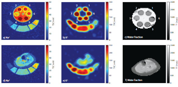
The conductivity and permittivity of tissue determine how electromagnetic fields are absorbed, but they differ between patients and are usually taken from literature tables. With TiCEPT (Tissue Composition based Electrical Properties Tomography) we estimate wideband electrical properties from tissue composition measured with quantitative multinuclear MRI of hydrogen, sodium and potassium. This links imaging directly to patient-specific modelling. See Model Inference & Calibration.
Imaging for Treatment Planning
Patient-specific treatment planning starts with an accurate 3D model of the patient. We developed automatic segmentation of head and neck CT and MR images, multimodal and groupwise image registration, and methods that adapt models to MR images acquired in treatment position.
Representative Publications
TiCEPT: Wideband Electrical Properties Tomography by Tissue Composition Assessment With Quantitative Multinuclear MRI
Barendsz LJC, Sumser K, Gerhalter T, Mestrom RMC, Nagel AM, Paulides MM · Magnetic Resonance in Medicine, 2026
DOI: 10.1002/mrm.70139
Clinical Performance and Future Potential of Magnetic Resonance Thermometry in Hyperthermia
Feddersen TV, …, Paulides MM · Cancers, 2021
DOI: 10.3390/cancers13010031
Magnetic Resonance Thermometry for Hyperthermia in the Oropharynx Region
Feddersen TV, …, Paulides MM, …, Poot DHJ · International Journal of Hyperthermia, 2024
DOI: 10.1080/02656736.2024.2352545
MR Thermometry Accuracy and Prospective Imaging-Based Patient Selection in MR-Guided Hyperthermia Treatment for Locally Advanced Cervical Cancer
VilasBoas-Ribeiro I, …, Paulides MM · Cancers, 2021
DOI: 10.3390/cancers13143503
Experimental Validation of the MRcollar: An MR Compatible Applicator for Deep Heating in the Head and Neck Region
Sumser K, …, Paulides MM · Cancers, 2021
DOI: 10.3390/cancers13225617
A Printed Yagi-Uda Antenna for Application in Magnetic Resonance Thermometry Guided Microwave Hyperthermia Applicators
Paulides MM, …, Smolders AB · Physics in Medicine & Biology, 2017
DOI: 10.1088/1361-6560/aa56b3
All selected publications
Frequently Asked Questions
What is MR thermometry?
MR thermometry uses magnetic resonance imaging to measure temperature changes in the body without inserting probes, most often based on the proton resonance frequency shift of water. It provides 3D temperature maps during thermal therapies such as hyperthermia.
What is electrical properties tomography?
Electrical properties tomography estimates the conductivity and permittivity of tissue from MRI. TiCEPT, developed in the EM4Care+Cure lab, derives these properties over a wide frequency range from tissue composition measured with multinuclear MRI.